X-RAY FINDINGS IN DIABETES FOOT MELLITUS SYNDROME
Abstract
Background: In 2020, 537 million adults were known to have been affected by diabetes, and one out of every ten individuals has diabetes mellitus. Diabetic foot syndrome is believed to affect 6.3% of individuals worldwide. It is the commonest cause of hospital admission and often leads to limb amputations. Radiology plays an essential role in early diagnosis for effective management and follow-up. Peripheral neuropathy, infections, and micro- and macrovascular pathways are common causes of these DMFS which have high morbidity and mortality rates.
Methodology: A cross-sectional retrospective study spanning two years on 90 adult diabetic patients with foot syndromes in a tertiary health care centre in South West Nigeria analysed demographic information, common sites, comorbidities, and X-ray findings using SPSS 23.0. Non-diabetic patients and those with non-diabetic foot syndrome were excluded.
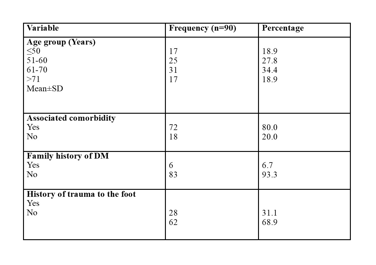
Results: Most participants were 61–70 years old (34.4%), and 80% had comorbidities. Only 6.7% had a positive family history of DM. 68.9% had no previous trauma before the onset of DMFS. The left foot was the most commonly affected (44.4%). Soft tissue swelling was the most common radiographic finding (15.6%). Calcaneal spurs, gangrene, and cellulitis were also seen in some participants. Osteopenia was a rare finding (1.1%).
Conclusion: X-rays are a good first imaging method, but a comprehensive diagnosis using multiple techniques is necessary for optimal patient care. Clinical evaluation can add value to X-ray imaging. This is important in resource-limited settings where financial constraints or fear of amputation may prevent seeking medical assistance.


